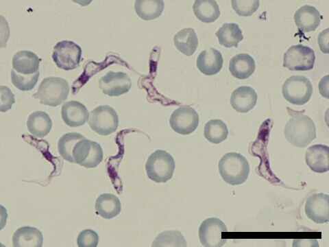
Blood stream form, trypomastigotes of Trypanosoma brucei brucei were obtained from peripheral blood of infected mouse. A nucleus and a kinetoplast were in the center and at the end , respectively. They were hardly stained with Giemsa’s solution. A flagellar base was near the kinetoplast. Flagellum was extended here with undulating membrane from the side of the cell body. The motility was shown by flagellar bending with flexion of the whole cell, so the bending form was observed. No clear morphological difference was found between T. b. brucei, and T. b. rhodesiense. This protozoan parasite causes animal trypanosomiasis (nagana) but is not pathogenic to humans. Bar indicated as 20 μm.