

|WHO研究協力センター|文部科学省COE拠点|GOARN研修|来たれ明日のウイルスハンター|
Home
研究テーマ・論文
WHO協力活動
大会・セミナー
人材募集案内
研究室紹介
これまでの記事
WHO COLLABORTING CENTRE REPORT (2002-2003)
1. Name of the Centre:
WHO Collaborating Centre for Reference and Research on Tropical Virus Diseases.
2. Address:
Department of Virology, Institute of Tropical Medicine, Nagasaki University, Sakamoto 1-12-4, Nagasaki City, Japan 852-8523.
3. Head of the Centre:
Kouichi Morita, Professor.
4. Terms of Reference:
1. to collaborate in epidemiological and virological studies on tropical
virus diseases;
2. to collaborate in the research and development of second generation
Japanese encephalitis vaccine, dengue virus vaccine and other tropical
virus diseases vaccines;
3. to collaborate in the development of rapid diagnostic methods of tropical
virus diseases;
4. to provide training on laboratory diagnosis and research on tropical
virus diseases;
5. to report the results of the studies to WHO and promote the exchange
of information on tropical virus diseases.
Place and date: Nagasaki, Japan, December 18th, 2003.
Signature: Kouichi Morita
Head of the Centre
5. Work performed in relation to the terms of reference:
1) Epidemiological and virological studies on tropical virus diseases:
1-1) Studies on serological surveillance of Japanese encephalitis virus infection in The Philippines.
Supported by the Grant-in-Aid for international Collaborative Research from the Ministry of Education, Science, Sports and Culture of Japanese Government, field studies were undertaken in collaboration with St. Luke's Medical Center and University of the Philippines. The objective of the first part of this study was to determine the impact of Japanese encephalitis among mild fever cases and severe encephalitis and/or meningitis cases. Seven hundred and seventy serum samples collected in 1999 from patients clinically diagnosed as dengue fever or dengue hemorrhagic fever, and 203 cerebrospinal fluid (CSF) and 50 sera collected from 1999 to 2002 from patients showing clinical encephalitis and/or meningitis, were examined using IgM capture ELISA for dengue and JE viruses. Out of 770 serum samples from patients with dengue fever or dengue hemorrhagic fever, 35% were positive for dengue, 0.8% were positive for JEV, 17% were double positive with dengue titer JEV titer, and 6% were double positive with JEV titer > dengue titer. Forty one percent of the samples were negative for either dengue or JE viruses. Analysis of samples from patients with encephalitis and/or meningitis showed that 7% of CSF and 24% of serum samples were IgM positive against JEV. Interestingly, for one fatal case both CSF and serum were positive for IgM against dengue virus. Summarizing, the results showed that 7% of cases were considered as JEV infection among mild fever cases and that the prevalence of JEV infection among severe encephalitis and/or meningitis cases was around 7-24%. The second part of this study aimed to monitor the seasonality of JEV infection in the Philippines. For that purpose, IgM and IgG antibody levels against JEV were monitored in pigs in Rizal province monthly for a period of one year (May 2002-April 2003). The result of IgG positive ratio against JEV indicated that the peak season of JEV infection was from July to September in 2002. Results of this serological surveillance on JEV indicated that JEV infection is widely prevalent among humans and pigs in the Philippines.
1-2) Distribution of three arbovirus antibodies among monkeys (Macaca fascicularis) in the Philippines.
Serum samples from 54 monkeys were collected from healthy individuals in a monkey farm in Luzon Island, Philippines, in 1999. Samples were examined by IgM-capture ELISA and also by indirect IgG ELISA for the presence of dengue, Japanese encephalitis (JEV) and chikungunya (CHIK) viruses. IgM ELISA showed a positive rate of 4%, 35% and 15% against dengue, JE and CHIK viruses, respectively. IgG ELISA showed higher positive rates, 100% against JE or dengue viruses and 59% against CHIK virus infections, among monkeys in the Philippines. The results indicated high prevalence of flavivirus infections and low prevalence of chikungunya virus infection, as well as suggested a possible sylvatic transmission cycle for these viruses.
2) Molecular biological studies of arboviruses.
2-1) Locus of a virus neutralization epitope on the Japanese encephalitis virus (JEV) envelope protein determined by use of long PCR-based region specific random mutagenesis.
Recombinant JEV populations possessing random mutations at the envelope (E) protein region were prepared by a long PCR method. Neutralization resistant mutants were selected by the used of a JE-specific virus neutralizing monoclonal antibody (mAb) 503. The mutants were classified into three groups, each containing two aminoacid changes at the E protein region, 52, Gln-Arg, and 136, Lys-Glu; 136, Lys-Glu, and 275 Ser-Pro; and 126 Ile-Thr, and 136, Lys-Glu, respectively. Furthermore, three different genetically engineered JEV variants containing a single mutation each (126 Ile-Thr, 136, Lys-Glu and 275 Ser-Pro) showed partial but not complete recovery of reactivity to mAb 503. These mutations were localized at the junction of domains I and II of the E protein. The results in these study indicated that substitution at aminoacid positions 52, 126, 136 and 275 altered the structure of the neutralization epitope for mAb 503 in the E protein of JEV. This study also demonstrated the efficacy of the long PCR-based recombinant virus technique as a useful tool for the creation of virus mutants carrying random mutations.
2-2) Complete nucleotide sequence of chikungunya virus and evidence for an internal polyadenylation site.
This study accomplished the complete nucleotide sequence of chikungunya
virus (S27 African prototype) and the presence of an internal polyadenylation
site was identified within the 3' non-translated region of this strain.
The complete genome of chikungunya virus S27 was 11, 805 nt in length;
excluding the 5' cap nt, the internal-poly adenylation (poly A) and the
3' poly A tail. It comprised two open reading frames, one encoding the
non-structural proteins (2474 aminoacids) and the other encoding the structural
proteins (1244 aminoacids). RNA secondary structures within the 5' non-translated
region (NTR) and repeated sequence elements (RSEs) within the 3' NTR,
were identified. In this study, secondary structures were predicted for
the 5¢ NTR of alphaviruses. The first stem-loop structure identified at
the 5¢ terminus (nt 2-26) of chikyngunya virus was identical to those
identified for Ross River and O'nyong-nyong viruses. The second stem-loop
structure (nt 32-71) was only identical to that found for Ross River virus.
O'nyong-nyong virus, however, possessed a separate loop in its second
stem-loop structure and the angulation was different. The 3¢ NTR of chikyngunya
virus contained three RSEs at positions 11382-11416, 11580-11614 and 11666-11701.
The secondary structures of the RSEs of chikungunya, Ross River and Barmah
Forest viruses were predicted using the first 24 nucleotides of each RSE.
All of the predicted hairpin structures were identical. Interestingly,
although O'nyong-nyong virus is closely related to chikungunya virus,
it did not contain any of the RSE found for the latter one. Chikungunya
virus isolate S27 used in this study, as well as the O'nyong-nyong isolate
(Levinson et al., 1990) manifested different types of RSEs and secondary
structures.
Phylogenetic analysis for the predicted aminoacid sequences of the compete
coding region, the non-structural polyprotein and the structural polyprotein
was carried out. Based on amino acid sequence homologies, phylogenetic
analysis of non-structural and structural proteins and characteristic
RSEs, this study concluded that chikungunya virus is closely related to
O'nyong-nyong virus, but is however a distinct virus. In addition, it
was confirmed the existence of an internal-polyA fragment which was found
to be of different lengths (39, 57, 68, 74, 77 and 84 adenine nucleotides)
but it started at an identical initiation position in different clones.
These results strongly suggested a capacity of the polymerase of alphaviruses
to create polyA by a template dependant mechanism.
3) Studies on cell death induced by flavivirus infection of cultured cells.
3-1) West Nile and St. Louis encephalitis viruses induced cytopathology.
In order to analyze the mechanism of cytopathology induced by the flaviviruses, experiments were carried out to observe whether apoptosis was induced in flaviviruses-infected cells. For this purpose, encephalitis viruses such as West Nile and Saint Louis encephalitis viruses were used to infect K562 (human mononuclear cell line) and Neuro 2a (N2a, mouse neuroblastoma cell line) cell lines. They were grown in RPMI 1640 or E-MEM medium supplemented with 10% heat-inactivated foetal calf serum at 37 °C in a 5% CO2 atmosphere. The cells were infected at MOI of 1 FFU/cells, and harvested at various times post infection. Analysis comprised various techniques. Cell viability was determined by the Trypan Blue exclusion test. The percentage of infected cells was determined by flow cytometry using the FITC-labelled anti-flavivirus group monoclonal antibody (FITC-6b6c-1). Cells undergoing apoptosis were recognised by the following methodologies: a) determination of DNA content by propidium iodide (PI) staining using hypotonic fluorochrome solution; b) detection of the characteristic translocation of phosphatidylserine to the outer cell membrane by the Annexin V-cy3 Apoptosis detection Kit; and c) identification of nuclear condensed chromatin by Hoechst staining. Results showed that infected K562 and Neuro 2a cells underwent apoptotic cell death, whereas cells inoculated with UV inactivated virus and mock-infected cells did not develop cytopathic effects. The gene expression of regulators of apoptosis bax, bcl-2 and bcl-xl was investigated. A rise in the expression of the pro-apoptotic bax gene was detected in the infected cells. These findings suggested that up-regulation of bax mRNA correlated with cytopathic effects observed in infected cells.
3-2) studies were carried out in order to determine whether human dendritic cells (DCs) are permissive to Japanese encephalitis virus (JEV) growth and to characterize JEV infection in these cells.
DCs were prepared by treating human monocytes (isolated by magnetic cell sorting using CD14 MicroBeads) with Interleukin-4 and Granulocyte-Macrophage Colony Stimulating Factor for 1 week. The confirmation of the appropriate phenotype of immature DCs (iDCs) was carried out by flow cytometry using phycoerythrin (PE) or fluorescein isothiocyanate (FITC) labeled anti-CD14, and -CD1a, antibodies. IDCs were infected with JEV JaOH0566 at MOI 1, 10 and 50 FFU/cell and harvested at various times after infection. The presence of JE viral infection was demonstrated by flow cytometry and Electron Microscopy. The highest percentage of infected cells was registered at 12 hs after infection. Production of replication competent viral particles was demonstrated by monitoring the JEV titer in the cell-free supernatant of JEV-infected iDCs. Titers ranged from 103 to 107 according to the MOI used and the time post infection. Morphological changes concomitant with cell death by apoptosis were observed in the JEV infected iDCs but not in the mock-infected iDCs. The occurrence of this phenomenon was confirmed by flow cytometry analysis using a single staining method for DNA content determination (propidium iodide (PI) hypotonic fluorochrome solution) and a double staining method to differentiate apoptosis from necrosis (FITC-Annexin V+ PI). Cells with lower DNA content reached 38% for JEV infected DCs at 48 hs after infection while mock-infected cells never showed more that 15%. Double staining experiments confirmed cell death by apoptosis. Results in this study indicated that JEV may initially infect and replicate in intraepithelial dendritic cells.
4) Studies on pathogenesis of dengue virus infection and dengue hemorrhagic fever.
4-1) Correlation between increased platelet-associated IgG and trombocytopenia in secondary dengue virus infections.
This study was done in collaboration with St. Luke's Medical Center and University of the Philippines, and supported by the Ministry of Education, Science and Culture of Japan and St. Luke's Medical Center.
In order to elucidate the mechanism of trombocytopenia in dengue hemorrhagic fever, the relationship between platelet-associated IgG (PAIgG) and platelet count in 53 patients in the acute phase of secondary dengue virus infection was investigated. A significant reverse correlation between the two parameters was found in the patients, while no correlation was observed in healthy volunteers. The low baseline platelet counts during the acute phase in 12 patients with secondary dengue virus infection significantly increased during the convalescent phase, while the increased PAIgG levels during the acute phase in these patients significantly decreased during the convalescent phase. The involvement of anti-dengue virus IgG was shown in platelets from 8 patients in the acute phase of dengue virus infection. The results of this study suggest that the formation of PAIgG involving anti-dengue virus IgG plays an important role in the induction of transient trombocytopenia observed in the acute phase of dengue virus infection.
4-2) In order to analyze the major replication sites(s) of dengue virus in the blood components of patients with dengue fever (DF) and dengue hemorrhagic fever/ dengue shock syndrome (DHF/DSS), flow cytometry experiments were carried out.
This study was conducted in Ho Chi Minh city, Vietnam, as part of a Japan-Vietnam Collaboration Project supported by the Japan Science Promotion Society (JSPS). Blood samples were collected from almost 100 clinically suspected dengue patients, and 17 were RT-PCR positive for dengue virus serotype 2. RT-PCR positive samples were analyzed by two-color flow cytometry using phycoerythrin (PE) labeled anti-CD3, -CD14, -CD16, or -CD19 antibodies and fluorescein isothiocyanate (FITC) labeled 6B6C-1, which is an anti-flavivirus group antibody. Out of 17 RT-PCR positive cases, one, a primary infection, showed the presence of B lymphocytes infected with dengue virus. Results in this study suggested that during primary dengue virus infection, the site of virus replication may be different and would involve B lymphocytes.
4-3) Single Arg-to-Ile mutation in the pre-membrane protein of dengue 2 virus associated with increased infectivity and enhanced cytokine production.
Previous genetic analyses indicated dengue serotype 2 strain-specific sequence differences that correlated well with disease severity. Amino acid residues Ile at premembrane 16 (PrM-16-Ile) and PrM-81-Thr were identified in dengue 2 strains isolated from Thai patients with dengue haemorrhagic fever (DHF) and dengue shock syndrome (DSS). Site-directed mutagenesis was used to investigate these two PrM loci. An infectious cDNA clone (pD2/IC-30P-A) of dengue 2 16681 virus, which contained the PrM-16-Arg and PrM-81-Thr, was used to construct a viral mutant Mut16 containing PrM-16-Ile. The PrM-81 Thr-to-Ala mutation was apparently lethal. For the variant Mut16, viral infectivity, replication and/or cytokine induction was investigated in human K562 cells, peripheral blood mononuclear cells, and immature myeloid dendritic cells (DCs). At 48 h after infection, the Mut16 virus showed a significantly higher level (68.7%) of infectivity in K562 cells, compared to the parental clone-derived D2/IC-30P-A virus (3.14%) and an engineered Mut16-revertant (PrM-16 Ile-to-Arg) virus (3.04%). Furthermore, the Mut16 virus infected 49.7% of DCs at 24 h post-infection, compared to 2.37 and 6.17% of cells infected with D2/IC-30P-A and Mut16-revertant viruses, respectively. Mut16 virus induced significantly higher levels of certain cytokines, including TNF-a, IL-6, and IL-12, compared to D2/IC-30P-A and Mut16-revertant viruses. These results suggested that the presence of the amino acid Ile at PrM-16 in DEN-2 virus may play a crucial role in the pathogenesis of DHF/DSS.
5) Laboratory diagnosis on arboviruses and other viruses.
5-1) Combined detection and genotyping of chikungunya virus by a specific RT-PCR.
The epidemiology and clinical features of chikungunya have a number
of similarities to those of dengue viruses. Both viruses are prevalent
in the tropics and subtropics and there is a possibility of large simultaneous
outbreaks involving these two viruses. Unlike dengue hemorrhagic fever
and dengue shock syndrome, chikungunya is rarely
fatal and patients and they not usually require hospitalization. Therefore,
it is very important to differentiate chikungunya infection from dengue
virus infection, especially in areas where dengue is endemic. A reverse
transcription-polymerase chain reaction (RT-PCR) was developed for the
detection of chikungunya virus infection. Two primer sets were designed
based on the non-structural protein 1 (nsP1) and the glycoprotein E1 (E1)
genes, which had well conserved nucleotide sequences among the same complex
of viruses but were specific to the 3' end of the chikungunya viral genome.
The expected lengths for the PCR products were 354 and 294 base pairs
for the nsP1 and E1 genes, respectively. RNA templates of dengue viruses
types 1 to 4 were all negative for both primer sets. The nsP1 primer set
was found to be more sensitive than the E1 primer set. These results suggested
that nsP1 primers would be suitable for diagnostic purposes, while the
E1 primers could be applied for genotyping of chikungunya virus strains.
5-2) Development of a real time- reverse transcription loop mediated isothermal amplification (RT-LAMP) for rapid detection of viruses.
5-2-1) Real time RT-LAMP for rapid detection of West Nile virus.
A one-step single tube real-time accelerated reverse transcription loop mediated isothermal amplification (RT-LAMP) assay was developed for detecting the envelope (E) gene of West Nile (WN) virus. The RT-LAMP assay is a novel method of gene amplification that amplifies the nucleic acid with high specificity, efficiency and rapidity under isothermal condition employing a set of six specially designed primers that recognize eight distinct sequences of the target. The whole procedure is very simple and rapid and the amplification can be obtained in less than one hour by incubating all the reagents in a single tube with reverse transcriptase and Bst DNA polymerase at 63°C. The detection of gene amplification could be accomplished by agarose gel electrophoresis as well as by real-time monitoring in an inexpensive turbidimeter. RT-LAMP assay was found to be 10 fold more sensitive than RT-PCR, and it had a detection limit of 0.1 PFU of virus. The specificity of the RT-LAMP assay was validated by observing no cross-reaction with other closely related members of the Flavivirus genus followed by restriction digestion and nucleotide sequencing of the amplified product. The results of this study indicated that RT-LAMP assay was rapid, cost effective, highly sensitive, specific and had a potential usefulness for rapid comprehensive WN virus surveillance.
5-2-2) Development and evaluation of a novel LAMP method for rapid detection of SARS Corona virus.
This study was conducted as part of a Japan-Vietnam Collaboration
Project supported by the Japan Science Promotion Society (JSPS).
A real-time quantitative RT-LAMP assay was developed in collaboration
with EIKEN Chemical Co. (Tokyo, Japan) and evaluated for rapid detection
of SARS-CoV rep gene. A total of 49 samples (15 throat washes, 13 throat
swabs, 21 combined throat & nasal swabs) collected from patients admitted
at the Hanoi-French and Ninhbinh hospitals, Vietnam during the SARS epidemic
were evaluated and compared to conventional RT-PCR. The RT-LAMP assay
showed 100 fold higher sensitivity and a detection limit of 0.01 PFU.
The assay procedure is simple as the amplification is carried out in a
single tube under isothermal conditions (63oC) and the result can be obtained
in less than 1 hr. Therefore, this RT-LAMP assay has the advantages of
rapid amplification, simple operation and easy detection. Furthermore,
it can be useful for rapid and reliable clinical diagnosis of SARS-CoV
in developing countries.
5-3) Serodiagnosis of Japanese encephalitis among Nepalese patients by the particle agglutination assay.
Serological diagnostic kits for routine diagnostic use in the southwestern region of Nepal, where Japanese encephalitis (JE) is a serious health problem, have not been available. This study was carried out to examine the applicability of a particle agglutination (PA) assay for JEV-IgM and to evaluate its accuracy for clinical diagnosis of JE. One hundred and ninety-three blood samples were collected from patients clinically diagnosed as having JE or other infectious diseases in the JE-endemic southwestern region of Nepal, in 2000. These samples were tested by PA assay, and JEV IgM-capture ELISA was used as a reference test system. Eighty-six samples were IgM-positive by the PA assay, and 71 of these 86 were also positive by IgM-capture ELISA. PA assay showed a sensitivity of 99% and a specificity of 88%. Results in this study indicated that PA assay was proved to be a simple, reliable and useful diagnostic test for JE diagnosis in rural hospitals.
6) Enzymatic activity of JE NS5.
The complete non-structural NS5 gene of Japanese encephalitis virus JaOH0566 strain was amplified and cloned into an expression vector. The NS5 protein was expressed in Escherichia coli and purified by His-taq based affinity chromatography. This recombinant NS5 protein exhibited RNA dependent RNA polymerase (RdRp) activity in vitro without any other viral or cellular factors. The RNA polymerase activity was dependent on the divalent cations. To better understand the mechanism of JEV genome replication, about 1 kilo-base RNAs corresponding to the 3' end of plus and minus strand RNA of JEV and dengue-2 virus were in vitro transcribed and used as templates for in vitro RNA synthesis catalyzed by the expressed JEV NS5 protein. It was found that JEV NS5 in vitro-expressed protein was more efficient in using the minus strand RNA as template. Serial deletions made on the JEV RNA templates demonstrated that the 3' end sequence of the JEV plus strand RNA has an inhibition effect on RNA synthesis. Using site directed mutagenesis analysis four aminoacid sequence motifs, important for RdRp activity, were identified. Substitutions in the conserved residues within these motifs led to a complete inactivation or severe impairment of enzyme activity.
7) Molecular epidemiology of Japanese encephalitis (JE) and dengue viruses.
7-1) Shift in JEV genotype circulating in northern Vietnam:
Implications for frequent introductions of JEV from Southeast Asia to
East Asia.
This study was conducted as part of a Japan-Vietnam Collaboration Project supported by the Japan Science Promotion Society (JSPS). Data on molecular epidemiology of JEV in Vietnam is very limited. Previously reported studies examined several JEV isolates from North and South Vietnam covering a time span of 27 years (1962-1988) through the capsid-prM region. All isolates analyzed in those studies belonged to genotype 3 suggesting that this was the main genotype circulating in Vietnam. In order to better understand the molecular epidemiology of JEV in Vietnam, the evolutionary relatedness of 16 JEV isolates (9 from Vietnam and 7 from Japan) to previously published JEV strains using E gene sequence data was examined. Vietnamese and Japanese strains isolated between 1986 and 1990 were found to cluster in genotype 3. However, more recent Vietnamese and Japanese strains isolated between 1995 and 2002, grouped within genotype 1, now a dominant, though previously unreported, genotype in Vietnam. In addition, strains isolated between 1995 and 2002 were closer related to those previously reported isolates from the '90s than to older genotype 1 strains. Considering that Japan and Korea also reported the introduction of genotype 1,the results in this study suggested that JEV genotype shift may be occurring throughout all East Asia. JEV has spread greatly during the past few decades, and migrating birds, particularly the black-crowned night heron and the Asiatic cattle egret are thought to be important in the dispersion of this virus to new geographic boundaries. Analysis of bird migration patterns along the East Asian-Australasian flyway indicated that spread by viremic migratory birds may provide a sound explanation to the widening distribution of JEV genotypes. This was further supported by results from phylogenetic analysis in this study in which not only for genotype 1 but also for genotype 3, temporally proximal isolates either from Vietnam, Korea or Japan were closely related among themselves. The results in this study on the spread of JEV to new territories suggest potential implications for future spread of West Nile virus in the case it reaches Southeast Asia; bird migration might contribute to its rapid dispersal throughout East Asia.
7-2) Molecular epidemiology of dengue virus serotype 4 in Vietnam.
This study was conducted as part of a Japan-Vietnam Collaboration Project
supported by the Japan Science Promotion Society (JSPS).
Dengue virus infection is a serious problem in Vietnam. Until 1998, the
major circulating serotypes were serotypes 1 and 2, but since 1997 srotype
3 appeared and in 1998 an outbreak of this serotype occurred. Dengue 4
cases were very few and a significant increased in the number of cases
was observed in recent years. In order to understand the molecular epidemiology
of dengue virus serotype 4 in Vietnam, 7 strains from southern Vietnam,
isolated between 1997 and 2001, were compared to 18 geographically distributed
previously published strains by phylogenetic analysis of 1,637 nt contained
within the membrane (M) and envelope (E) genes and the M-E junction region.
Previous studies showed that dengue virus serotype 4 isolates could be
grouped into two genotypes (G), named G1 and G2. Isolates in this study
clustered together in a separate cluster which was assigned as G3.
These southern Vietnamese dengue serotype 4 isolates presented three aminoacid
changes in the E gene, unique to the new assigned genotype 3. They are
Met at position 96, Glu at position 210 and Thr at position 253.
7-3) Molecular epidemiology of dengue virus serotype 3 in Bangladesh.
This study was supported by the Japan Society for Technical Cooperation (JST)- CREST. Bangladesh first experienced a dengue virus outbreak in the year 2000. The main virus serotype circulating was identified as serotype 3, but virus isolation was not successful. In order to study the molecular epidemiology of the 2002 dengue virus outbreak in Bangladesh, dengue virus was isolated in C6/36 cell line, purified and the envelope (E) gene of 8 isolates sequenced. The evolutionary relatedness of these 8 Bangladeshi isolates to a large group of previously published strains was carried out. All isolates in this study clustered within genotype 2, but they were grouped in a distinct sub-cluster of this genotype.
8) Training on laboratory diagnosis and research on tropical virus diseases:
8-1) Training Course on Tropical Medicine:
This course was organized from June 1st to August 31st consecutively
during the years 2000, 2001, 2002 and 2003. Japanese participants received
training in
virus isolation, serological assays (IgM capture ELISA) and RT-
PCR. In year 2003 a demonstration of Loop Mediated Isothermal
Amplification (LAMP) was also given. This training took place in the Virology
Department during the following periods:
- August 16th-22nd, 2000.
- August 7th-14th, 2001.
- August 5th-13th, 2002.
- August 4th - 8th, 2003.
8-2) Training overseas students:
Participants supported by the Japan International Cooperation
Agency (JICA).
1- Ms. Pooi Fong Wong from the Department of Medical Microbiology, Faculty
of Medicine, University of Malaya, Malaysia, from January 10th to December
18th 2000, conducted molecular biological manipulation of RNA for the
construction of chimera viruses.
2- Mr. Kriengsak Limkittikul from the Department of Pediatrics, Tropical
Medicine Faculty, University of Mahidol, Thailand, from from January 10th
to December 17th 2001, conducted studies on effect of random mutations
introduced by PCR in the replication of JEV.
3- Ms. Gissel Garcia Menendez from the Department of Virology, Laboratory
of Immunology, Institute Pedro Curi, Cuba, from from January 10th to December
18th 2002, studied and characterized JEV infected dendritic cells.
4- Dr. Guillermo Posadas Herrera from the Technological Institute of Madero
City, Mexico, from from January 10th to December 13th 2003, developed
a RT-LAMP assay for the detection of the envelope gene of West Nile virus.
8-3) Ph.D. Graduate students in Medical Sciences in Nagasaki University, supported by Monbusho Scholarship from the Ministry of Education, Science, Sports and Culture of Japan.
8Mr Afjal Hossain Khan, from Bangladesh since 1998, successfully
defended his thesis on "Complete nucleotide sequence of
chikungunya virus and evidence for an internal polyadenylation
site", and received his Ph.D. degree in March 2003.
Mr. Edward Gitau M. Mathenge, from Kenya since 1999.
Mr. Yu Fu Xun, from China since 2001.
Ms. Thai Hong Thicam, from Vietnam since 2001.
Ms. Leonora Salda, from The Philippines since 2001.
8-4) Non Degree Research Fellow supported by Monbusho Scholarship from the Ministry of Education, Science, Sports and Culture of Japan.
Dr. Manmohan Parida from the Department of Virology,
Defence Research and Development Establishment, Ministry of
Defence, Government of India, since October 2002, and developed
a RT-LAMP assay for the detection of the envelope gene of West
Nile virus.
8-5) Rompaku fellow supported by the Japan Science Promotion Society (JSPS).
Mr. Paresh Sumatilal Shah, Research Officer, National Institute
of Virology, Poona, India, stayed for a period of three months in
the years 2000, 2001, 2002 and 2003, and carried out studies on
JEV DNA vaccine and effects on neuroinvasiveness of mutations
in the pr-M gene of Japanese encephalitis virus.
8-6) Fellows supported by the Japan Science Promotion Society (JSPS) as part of a Japan-Vietnam Collaboration Project.
Year 2000: From the Virology Department of the National Institute of Hygiene and Epidemiology (NIHE), Hanoi, Vietnam: Dr. Phan Thi Nga (1 month)
Year 2001: From the Virology Department of the National
Institute of Hygiene and Epidemiology (NIHE),
Hanoi, Vietnam:
Dr. Le Thi Quyen Mai (1 month)
Professor Truong Uyen Ninh (1 month)
From the Department of Virology of the Pasteur
Institute Ho Chi Minh City, Vietnam:
Dr. Vu Thi Que Huong (1 month)
Professor Do Quang Ha (1 week)
Year 2002: From the Virology Department of the National
Institute of Hygiene and Epidemiology (NIHE),
Hanoi, Vietnam:
Dr. Le Thi Quyen Mai (1 month)
Mr. Vuong Duc Cuong (1 month)
From the Department of Virology of the Pasteur
Institute Ho Chi Minh City, Vietnam:
Dr. Vu Thi Que Huong (1 month)
Year 2003: From the Virology Department of the National
Institute of Hygiene and Epidemiology (NIHE),
Hanoi, Vietnam:
Dr. Le Thi Quyen Mai (1 month)
Mr. Vuong Duc Cuong (2 month)
Mrs. Nguyen Thu Thuy (1 month)
Mrs. Vu Hong Nga (1 month)
From the Department of Virology of the Pasteur
Institute Ho Chi Minh City, Vietnam:
Mrs. Huynh Thi Kim Loan (1 month)
8-7) International Atomic Energy Agency (IAEA) fellows to be train in viral diagnostic of Dengue and Japanese encephalitis viruses.
Ms. Menaca Hopugoda from Sri Lanka, visited this department
for the period of three months in 2002.
Dr. Wimaladharma Abeyewickreme from Sri Lanka, visited this
department for the period of two weeks in 2003.
8-8) Fellows supported by the Human Science Foundation.
Dr. T. N. Dhole from the Department of Microbiology, Sanjay
Gandhi Post Graduate Institute of Medical Sciences, Lucknow,
India, visited this department for the period of two weeks in
2002.
8-9) Fellowship supported by the Japan Society for Technical Cooperation (JST).
Dr. Mohammend Alimul Islam from the Department of
Microbiology and Hygiene, Bangladesh Agricultural University,
Mymensingh, Bangladesh, since May 2002, is carrying studies
on molecular epidemiology of dengue virus in Bangladesh.
9) Collaboration between the Centre and WHO.
A global outbreak of severe acute respiratory syndrome (SARS) was associated with a novel corona virus, SARS-CoV. Transmission within hospitals was a major contributor to disease spread and amplification. Members of the Institute of Tropical Medicine, Nagasaki University were recruited by WHO to collaborate with their professional expertise.
Dr. Koichi Morita, Professor of the Department of Virology, was recruited,
from 3 to 17 April 2003, to investigate and supervise the preparation
of hospitals for SARS suspected patient's admission and isolation. Dr.
Morita visited The Philippines, Cambodia, Laos and Macao.
Dr. Kazunori Oishi and Dr. Hiroshi Watanabe from the Department of Internal
Medicine, were recruited from May 10 to June 11 and May 2 to June 15,
2003, respectively, to visit China.
11) Evaluation
The terms of reference of this Collaborating Centre are closely related to our research activities, with particular emphasis on arthropod-borne virus infections, such as Japanese encephalitis, dengue fever and dengue haemorrhagic fever, West Nile fever, chikungunya virus infection, etc. The designation of our WHO Collaborating Centre has absolutely promoted and enhanced its activities including exchange and training of researchers from developing countries in tropics. Epidemiological information and research developments have been disseminated through many ways, i.e. international publication, scientific congress, official public health meetings and internet homepage. Through these activities, our collaborating centre contributed a lot on capacity building of human resources in the Region and increasing awareness from governments and public on the importance of communicable disease research. Now the government of Japan is increasing its support for communicable disease response and research and our Centre received five post-doctoral posts from governmental research foundations, such as Japan Society for the Promotion of Science (JSPS), Japan Health Science Foundation (JHSF) and Japan Science and Technology Agency (JST). This certainly increased research capacity of the Centre and also enabled the Centre to dispatch tree expert to the WHO SARS Response Team in the Western Pacific Region during the SARS epidemic from April to June 2003. We hope that we can continue and fulfil our contribution in the research, training and response in the tropical and emerging virus infections by providing expertise and manpower as a WHO Collaborating Centre.
12) Scientific publications.
1) Parida M., Posadas M., Inoue S., Hasebe F., and Morita K.. Real-time
reverse transcription loop mediated isothermal amplification for rapid
detection of West Nile virus. J.Clin.Microbiol. 2004 (in press).
2) Morita K. Severe Acute Respiratory Syndrome (SARS). The Healthy Children. Vol. 374:42-43,2004.
3) Pandey B., Yamamoto A., Morita K., Kurosawa Y., Rai S., Adhikari S., Kandel P., and Kurane I.: Serodiagnosis of Japanese encephalitis among Nepalese patients by the particle agglutination assay. Epidemiol.Infect. 131:881-885,2003.
4) Morita K., Parida M. Mathenge EGM: Molecular basis of flavivirus pathogenesis. Cell Technology. 22:1178-1181:2003
5) Morita K.: West Nile virus infections. The Journal of Adult Diseases Vol.33: 1121-1124 2003.
6) Morita K.: Vaccination plan for Japanese encephalitis. Japan Medical Journal. Vol.4135: 145-146, 2003.
7) Morita K.: Arthropod borne virus: West Nile virus infection. The Healthy Children. Vol. 369:42-43,2003.
8) Morita K.: West Nile virus encephalitis and Japanese encephalitis. Clinical Microbiology. 30:351-355, 2003.
9) Ohishi K., Inoue S., Cinco M., Diaano E., Alera M., Alfon J., Abanes F., Crus D., Matias R., Matsuura H., Hasebe F., Tanimura S., Kumatori A., Moirta K., Natividad F., Nagatake T: Correlation between increased platelet-associated IgG and thrombocytopenia in secondary dengue virus infections. J.Med.Virol. 71:259-264, 2003.
10) Morita K.: Response for the outbreak of West Nile fever and encephalitis、Japan Medical Journal. Vol.4122: 101, 2003.
11) Inoue S., Morita K., Matias R.R., Tuplano J.V., Resuello R.R.G., Candelario J.R., Cruz D.J.M., Mapua C.A., Hasebe F., Igarashi A. and Natividad F.F. Distribution of three arbovirus antibodies among monkeys (Macaca facscicularis) in the Philippines. J.Med.Primatol. 32:89-94, 2003.
12) Morita K.: Dengue fever and dengue haemorrhagic fever. Nihon Rinsho. Vol.61:302-305,2003.
13) Morita K.: Prevention of West Nile fever. Journal of The Japan Pharmaceutical Association. Vol.55:73-75,2003.
14) Morita K.: Japanese encephalitis, diagnosis and treatment. Clinical Al-Round. Vol.52:1121-1125, 2003.
15) Parquet M.C., Kumatori A., Hasebe F., Edward G.M. Mathenge E.G.M. and Morita K.: Saint Louis Encephalitis Virus induced pathology in culture cells. Archives of Virology. Vol.147:1105-1119. 2002.
16) Pandey B.D., Thapa L.B., Sherchand J.B., Rimal N., Bhattarai A., and Morita K.: Etiology of diarrhoea among adult patients during the early monsoon period in Kathmandu, Nepal. J.J.Trop.Med.and Hygiene. Vol.30:133-137. 2002.
17) Afjal Hossain Khan, Kouichi Morita, Maria del Carmen Parquet, Futoshi Hasebe, Edward G. M. Mathenge and Akira Igarash..: Complete Nucleotide Sequence of Chikungunya Virus and Evidence of an Internal Polyadenylation site. J.Gen.Virol Vol.83:3075-3084, 2002.
18) Morita K.: Nipah virus infection. The Journal of Pediatric Practice. Vol.65:2117-2120, 2002.
19) Morita K.: Japanese encephalitis. Japanese Journal of Pediatric Medicine. Vol.34:1084-1086,2002.
20) Vargas R.E.M., Morita K., Eshita Y., Tsuda Y., Fukuma T. and Takagi M. Infection and dissemination of two dengue type 2 viruses isolated from patients exhibiting different disease severity in orally infected Aedes aegypti from different geographic origin. Journal of Medical Entomology and Zoology Vol.53:21-27. 2002.
21) Hasebe F., Parquet M.C., Pandey B. D, Mathenge E.G.M., Morita K., V. Balasubramaniam V., Saat Z., Yusop A., Sinniah, M., Natkunam S. and Igarashi A.: Combined Detection and Genotyping of Chikungunya Virus by a Specific Reverse Transcription-Polymerase Chain Reaction. J.Med.Virol. Vol.67:370-374. 2002.
22) Morita K.: Dengue vaccines. Clinical Microbiology. Vol.29:137-141, 2002.
23) Masaki H., Hasebe F., Ahmed K., Fukuda T., Furumoto A., Watanabe K., Oishi K., Igarashi K. and Nagatake T. A clinical, serological, and immunological study in a Japanese traveler with dengue fever. J Infect Chemother Vol.8:365-367, 2002.
24) Kamimura K., Horio M., Nakamura M., Doi R., Takashima I., Igarashi A., Ahmed A., and Takasu T.: Med Entomol. Zool. Sampling of Culex tritaemiorhynchus for virus isolation in Karachi and Heleji Lake, a suburb of Karachi, Pakistan. Med.Entomol.Zool.53:suppl.2:43-46. 2002.
25) Igarashi A.: Control of Japanese Encephalitis in Japan: Immunization of Humans and Animals, and Vector Control. in Japanese encephalitis and West Nile Virus. Current Topics in Microbiology and Immunology. Vol.267:139-152, 2002.
13) Presentations at International and Overseas Conferences.
1) Hasebe F., Limkittikul K., Hong T.C.T, Utama A., Shimizu H., Miyamura T., and Morita K.: Significance of mutant helicase motif II for Japanese encephalitis virus infectivity. Thiry-sixth Joint Working Conference on Viral Diseases. US/Japan Cooperative Medical Science Meeting. July 16-18, 2002. Matsumoto, Japan.
2) Morita K.: New technology for laboratory diagnosis on JE. SEAMIC Meeting, Oct 21-25, 2002. Manila, Philippines.
3) Morita K.: Research on new JE vaccines. SEAMIC Meeting, Oct 21-25, 2002. Manila, Philippines.
4) Morita K.: Virological research, new information on JEV. SEAMIC Meeting, Oct 21-25, 2002. Manila, Philippines.
5) Morita K, Wong PF, Methenge EGM, Kashiwaki T, and Hasebe F.: Construction and characterization of a Japanese encephalitis/dengue 2 chimeric virus. XII International Congress of Virology. July 27-Aug 1, Paris, France.
6) Morita K. Construction and characterization of Japanese encephalitis/dengue chimeric viruses Japan-Singapore International Symposium. Oct 10-11, 2002. Nagasaki, Japan
7) Morita K.: Dengue /JE chimeric viruses and their characterization. Japan-Vietnam International Symposium. Nov.28-30, 202. Hanoi, Vietnam.
8) Hasebe F., Limkittikul K., Hong T.C.T, Utama A., Shimizu H., Miyamura T., and Morita K.: Significance of mutant helicase motif II for Japanese encephalitis virus infectivity Japan-Vietnam International Symposium. Nov.28-30, 2002. Hanoi, Vietnam
9) Morita K.Pandey B., Kinney RM., Kumatori A., Hasebe F., Parquet MC. Inoue S., and Igarashi A: Single Arg-to-Ile mutation in the PrM Protein of dengeu 2 virus associated with inversed infectivity and enhanced cytokine production in human dendritc cells. 37th US-Japan Cooperative Medical Science Program. Houston, USA July 18-20, 2003
10) S. Inoue, MT Alonzo, DJM Cruz, F Tadena1, RR Matias, K. Morita and FF Natividad..Serological Surveillance of Japanese encephalitis virus infection in the Philippines. 15th Annual Convention of The Philippine Academic Society for Microbiology and Parasitology, Manila, Philippines. December 6, 2003.
11) Parida M., Posadas G., Inoue S., Hasebe F. and Morita K. Real-time reverse transcription Loop Mediated isothernal amplification for rapid detection of West Nile. 6th Asia Pacific Congress of Medical Virology, Kuala Lumpur, Malaysia December 7-10, 2003.
12) MT Alonzo, S. Inoue, DJM Cruz, F Tadena, RR Matias, K. Morita and Natividad FF Serological Surveillance of Japanese encephalitis virus infection in the Philippines. 6th Asia Pacific Congress of Medical Virology, Kuala Lumpur, Malaysia December 7-10, 2003.
13) Inoue S., Alonzo MT, Cruz DJM, Tadena F, Matias RR, Morita K. and Natividad FF. Serological Surveillance of Japanese encephalitis virus infection in the Philippines. 8th International Conference on Tropical Pathology, Quezon City, Philippines, December 12, 2003.
14) Morita K., Recent development of dengue research in WHO Collaborating Centre in Nagasaki University. WHO meeting for DengueNet Implementation in the Asia-Pacific Regions. Kuala Lumpur, Malaysia December, 11-13, 2003.
前のページへ戻る。世界保健機関西太平洋地域事務局
WHOはどんな機関?
The World Health Organization,
 the
United Nations specialized agency for health, was established on 7 April
1948. WHO's objective, as set out in its Constitution, is the attainment
by all peoples of the highest possible level of health. Health is defined
in WHO's Constitution as a state of complete physical, mental and social
well-being and not merely the absence of disease or infirmity.
the
United Nations specialized agency for health, was established on 7 April
1948. WHO's objective, as set out in its Constitution, is the attainment
by all peoples of the highest possible level of health. Health is defined
in WHO's Constitution as a state of complete physical, mental and social
well-being and not merely the absence of disease or infirmity.
WHO is governed by 192 Member States through the World Health Assembly. The Health Assembly is composed of representatives from WHO's Member States. The main tasks of the World Health Assembly are to approve the WHO programme and the budget for the following biennium and to decide major policy questions.
WHO研究協力センターの役割は?
Collaborating Center appointment by WHO indicates that an organization contributes to WHO's public health mission by providing specialized knowledge, expertise, and support in the health field to WHO and its member nations. Terms of reference, agreed upon by WHO and the Collaborating Center, define the role and responsibilities of a center.
ホームページに関するご意見、ご要望は、ホームページ担当窓口までお寄せください。
Denguenet Meeting
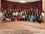
Denguenet Meeting, Kuala Lumpur, Malaysia. (平成15年12月11日から13日)
Institute of tropical medicine,
Nagasaki university.